You are not currently logged in. Please create an account or log in to view the full course.
A Review of the Skeletal System
- About
- Transcript
- Cite
The Musculoskeletal System
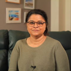
In this course, Dr Rina Pandya (University of the West of England) explores the musculoskeletal system. In the first lecture, we think about the human skeletal system and the key points of interaction within it. In an extension of the first lecture, we review the human skeletal system in more depth. In the second lecture, we think about the hip joint, femur and pelvis. In the third lecture, we think about bone shapes and types. Next, we think about muscle functions and methods of identification. In the fifth and final lecture, we think about types of joints and their key components.
A Review of the Skeletal System
In this lecture, we review the human skeletal system, focusing in particular on: (i) defining posterior as facing to the back, anterior as facing to the front, medial as facing to the centre, lateral as facing towards the outside, superior as facing towards the head and inferior as facing towards the feet; (ii) the primary function of the cranium being to protect the brain; (iii) the base of the skull, which is hollow to allow the spinal cord, arteries and veins to reach the brain; (iv) a review of the 33 vertebrae in the human spine, with their separation into cervical, thoracic and lumbar vertebrae, as well as the sacrum and coccyx; (v) the intervertebral disc as the presence defining whether vertebrae are defined as fused or separate; (vi) the three bones which make up the sternum being the manubrium, sternum body and xiphoid bones; (vii) the two clavicles, known as the collar bones, which join the sternum at the sternoclavicular joints; (viii) the role of the clavicles being to keep the chest open, with their omission from the skeletons of animals like cats enabling them to compress their chest to squeeze through small gaps; (ix) the role of the scapula providing a glenoid cavity, connecting with the head of the humerus to form the shoulder joint; (x) the radius on the outer edge and the ulna on the inner edge of the lower half of the arm; (xi) the outer and inner edges being defined by the default skeletal position of standing facing forwards with the palms turned forwards; (xii) the primary bones in the leg being the femur in the upper thigh, tibia in the lower leg, joined by the floating patella, or knee cap; (xiii) the fibula as the smaller, secondary bone in the lower leg; (xiv) the talus, which forms the ankle joint with the fibula and tibia; (xv) the metatarsals in the foot, with the fourteen phalange toe bones; (xvi) the seven cervical vertebrae below the base of the skull which, like all the vertebrae in the spine, have a hole through them for the spinal cord; (xvii) the role of the intervertebral discs as ‘shock absorbers’ between the vertebrae; (xviii) the meninges, which are the three layers forming the protection of the spinal cord; (xix) the first of the cervical vertebrae, C1, which is referred to as Atlas, named after the Green God who holds up the world.
Cite this Lecture
APA style
Pandya, R. (2024, December 17). The Musculoskeletal System - A Review of the Skeletal System [Video]. MASSOLIT. https://massolit.io/courses/the-musculosketal-system
MLA style
Pandya, R. "The Musculoskeletal System – A Review of the Skeletal System." MASSOLIT, uploaded by MASSOLIT, 17 Dec 2024, https://massolit.io/courses/the-musculosketal-system

